this all our services that we have mentioned down below

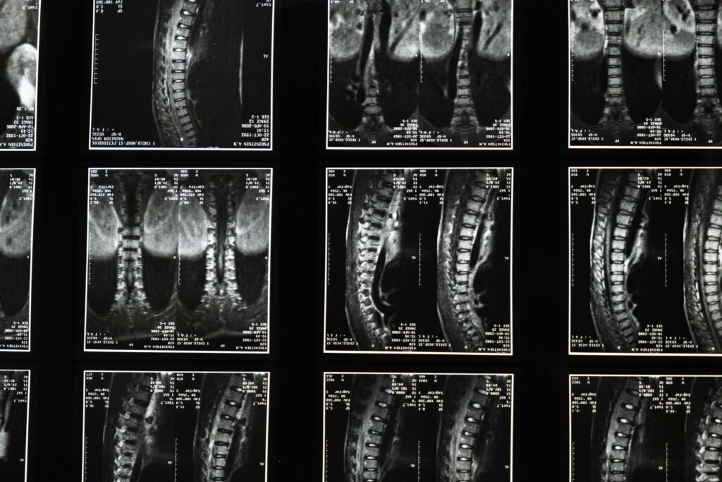
1>CT Scan
A computed tomography (CT) scan is a sophisticated imaging technique that utilizes X-ray technology to create detailed cross-sectional images of the body. This method allows healthcare professionals to visualize internal structures with remarkable clarity, aiding in the diagnosis and evaluation of various medical conditions. By combining multiple X-ray images taken from different angles, a CT scan generates comprehensive three-dimensional representations, which can be crucial for identifying tumors, internal injuries, or other abnormalities. The procedure for undergoing a CT scan is typically straightforward and non-invasive. Patients are usually required to lie on a motorized table that moves through a large, doughnut-shaped machine. During the scan, the machine rotates around the patient, capturing a series of images that are then processed by a computer to produce the final images. Depending on the area being examined, a contrast dye may be administered to enhance the visibility of certain tissues or blood vessels, providing even more detailed information for the interpreting physician. CT scans are widely used in various medical fields, including oncology, cardiology, and emergency medicine, due to their ability to provide rapid and accurate assessments. While the benefits of CT imaging are significant, it is essential to consider the associated risks, particularly the exposure to ionizing radiation. Therefore, healthcare providers carefully evaluate the necessity of a CT scan against potential risks, ensuring that patients receive the most appropriate and effective diagnostic care.
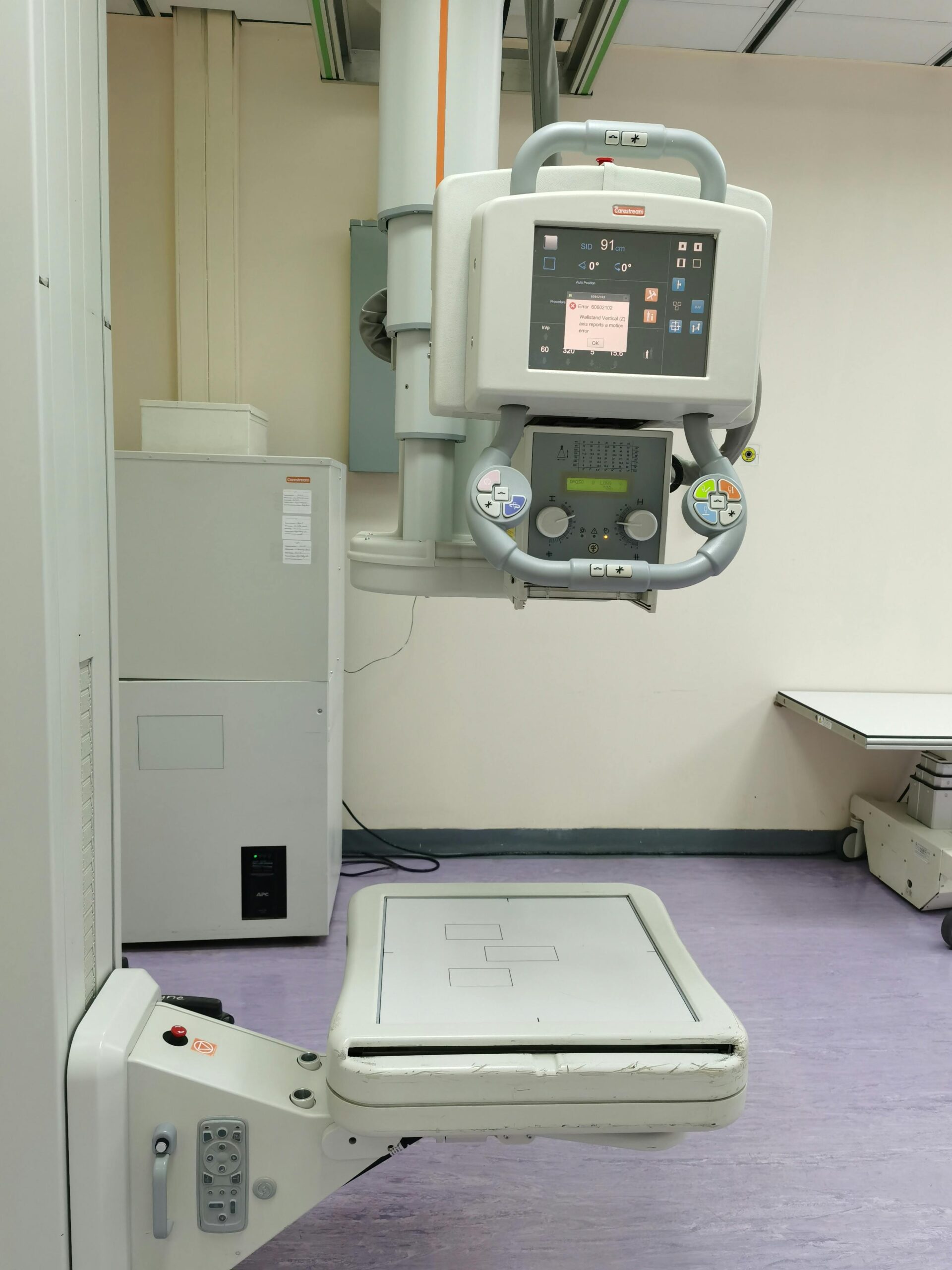
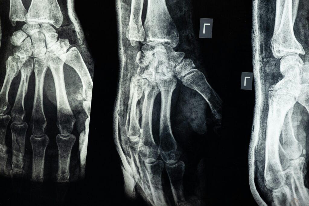
2>X-ray
X-ray imaging is a diagnostic tool that utilizes electromagnetic radiation to create images of the internal structures of the body. This non-invasive technique allows healthcare professionals to visualize bones, organs, and tissues, aiding in the detection and diagnosis of various medical conditions. By passing X-rays through the body, denser materials such as bones absorb more radiation, resulting in a contrast that highlights different anatomical features on the resulting images. The process of obtaining an X-ray is relatively quick and straightforward. Patients are typically positioned in front of an X-ray machine, and the technician will take images from various angles to ensure comprehensive coverage of the area of interest. While the procedure is generally safe, it is essential to minimize exposure to radiation, particularly for vulnerable populations such as pregnant women and children. Therefore, healthcare providers carefully assess the necessity of the X-ray in relation to the potential risks involved. In addition to its diagnostic capabilities, X-ray technology has evolved significantly over the years, leading to advancements such as digital X-rays, which offer enhanced image quality and reduced radiation exposure. These innovations have improved the accuracy of diagnoses and the overall efficiency of medical imaging. As a result, X-rays continue to play a crucial role in modern medicine, providing invaluable insights that guide treatment decisions and improve patient outcomes.
Road to success is always under construction